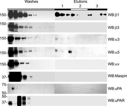
FIGURE 2.
Maspin affinity chromatography. HT1080 cell lysate was applied to a maspin affinity column, after incubation the column was washed extensively with PBS to remove unbound protein (Washes). Specifically bound proteins were then eluted by sequential application of 3 ml of PBS containing 0.5 m NaCl, 1.0 m NaCl, and acetate buffer pH 4.0, and collected as 1-ml fractions (Elutions, labeled 1–3, respectively). 30 μl of each fraction was separated by SDS-PAGE prior to Western blotting with primary antibodies to β1 (10 μg/ml), β3 (10 μg/ml), α3 (1:1000 antiserum), α5 (1:1000 antiserum), αv (10 μg/ml), maspin (1 μg/ml), uPA (10 μg/ml), or uPAR (10 μg/ml).

