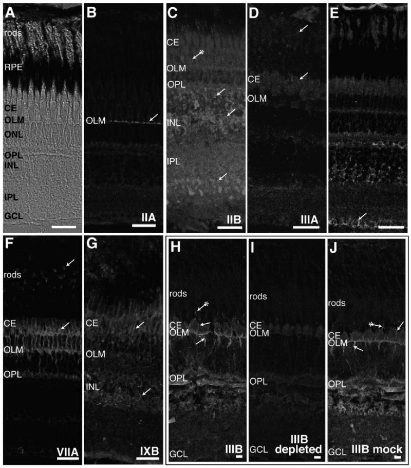
Figure 3.

Immunohistochemistry of adult fish retina with antibodies to different classes of myosins. A: The laminar arrangement of the adult zebrafish retina is seen with Nomarski optics. Light-adapted zebrafish retinal cryosections were stained with antibodies specific to myosins IIA (B), IIB (C), IIIA (D), VI (E), VIIA (F), and IXB (G). Light-adapted striped bass retinal sections were used for myosin IIIB immunostaining (H–J). B: Myosin IIA antibody stains only the outer limiting membrane (OLM) formed by termini of Müller cell processes. C: Myosin IIB antibodies brightly label cell bodies in the stratum containing predominantly bipolar cells in the inner nuclear layer (INL) and termini of cell processes found in the inner plexiform layer (IPL) and at the outer plexiform layer (OPL). Fluorescence can sometimes be seen in cone accessory segments with myosin IIB antibodies (indicated by an arrow with an attached asterisk). D: Rod inner segments and the distal region of cone ellipsoids are labeled with myosin IIIA antibodies. E: Staining with myosin VI antibodies is found in horizontal cells in the INL and ganglion cells (GCL). Some myosin VI immunostaining is also exhibited in the OLM and OPL. F: Myosin VIIA antibody fluorescence is found throughout cone inner segments and in rod inner segments. In cones, myosin VIIA staining is also located in accessory outer segments, axons, and synapses. Weaker labeling of myosin VIIA occurs in horizontal, amacrine, and ganglion cells. G: Myosin IXB is expressed in cone ellipsoids and accessory outer segments and in the amacrine cell region of the INL. H: Myosin IIIB antibody stains cone accessory outer segments, inner segments, and axons. Additional myosin IIIB fluorescence is localized to the OLM, OPL, and INL. I: Greatly reduced staining is observed when myosin IIIB antibody was preincubated with a fusion protein containing the myosin IIIB tail prior to incubation with striped bass retinal sections. J: Preincubation of myosin IIIB antibodies with a control fascin 2B fusion protein does not change the staining pattern in striped bass retina. RPE, retinal pigmented epithelium; CE, cone ellipsoid. Scale bar = 20 μm in A–J.
