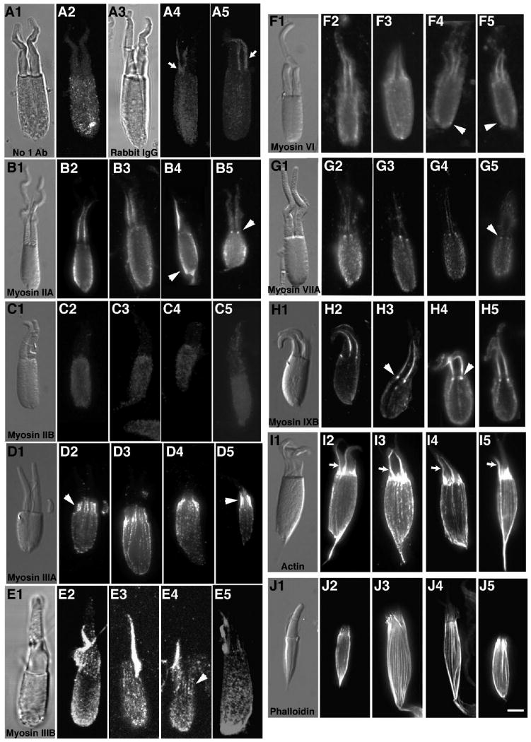
Figure 5.

Cone inner/outer segment preparations stained with antibodies to different classes of myosins. Preparations consisting of cone inner segment ellipsoids and attached outer segments from green sunfish were immunostained for various myosin family members and actin. A2: Faint background staining in the cone ellipsoid with the fluorescently tagged secondary antibody was observed in controls incubated without primary antibody. A4,5: Substitution of rabbit IgG for primary antibodies gave some nonspecific staining in the cone ellipsoid but resulted in brighter fluorescence located in the accessory outer segment, which was also observed in preps treated with other antibodies (indicated by small arrows). B2–5: Myosin IIA antibodies stained the ellipsoid surface membranes and basal bodies (arrowheads). C2–5: Myosin IIB antibody staining was the same as controls (i.e., low-level stain). D2–5: Myosin IIIA localized in a distinct pattern to the distal region of inner segment actin filaments, extending into the calycal processes surrounding the proximal outer segment (arrowheads). E2–5: Like myosin IIIA, some actin filament bundle staining in the calycal processes is exhibited near the proximal outer segment with myosin IIIB antibody (arrowhead), but there is additional staining of the ellipsoid. F2–5: Myosin VI antibodies stain the cone ellipsoids (arrowheads). G2–5: Myosin VIIA immunostains the two basal bodies found in double cones (arrowheads). H2–5: Myosin IXB antibodies also stain the basal bodies at the base of the outer segment (arrowheads). A monoclonal actin antibody (I2–5) and phalloidin (J2–5) brightly stain the actin filament bundles found running the length of the ellipsoid and terminating near the proximal end of the outer segments in the calycal processes. Scale bar = 5 μm in J5 (applies to A1–J5).
