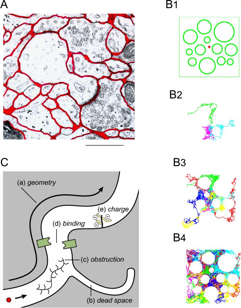
Fig. 1.
Basic concepts of ECS. A. Electronmicrograph of small region of rat cortex with dendritic spine and synapse. The ECS is outlined in red; it has a well-connected foam-like structure formed from the interstices of simple convex cell surfaces. Even though the ECS is probably reduced in width due to fixation procedure it is still evident that it is not completely uniform in width. Calibration bar approximately 1 μm. (Modified from Ref. 264). B1-B4. Molecules executing Random walks reveal structure. B1: cross-section through an idealized 2D square brain region bounded by impermeable walls and containing a number of circular cellular profiles. The ECS width is exaggerated. B2: Three molecules (different colors) have been released from the location marked with a red “+” in B1 and allowed to execute up to 150 random steps. If the molecules encounter a cell boundary or wall, the step is canceled and the next random step selected. The profiles of the cells are omitted from this and the two subsequent panels, only the trajectories of the random walks are shown. The three trajectories in B2 appear to have a random distribution. B3: 12 molecules execute random walks and now the aggregate of their trajectories begins to reveal the presence of the cells. B4: 48 random walks reveal an increasingly accurate view of the boundaries of the cells and the boundaries of the region. Note that the individual steps are large in this simulation to reduce the number required to reveal the geometry (Modified from Ref. 264). C. Factors affecting the diffusion of a molecule in the ECS. These are: a) geometry of ECS which imposes an additional delay on a diffusing molecule compared to a free medium. b) dead-space microdomain where molecules lose time exploring a dead-end. Such a microdomain may be in the form of a ‘pocket’ as shown but it may also take the form of glial wrapping or even a local enlargement of the ECS. c) Obstruction in the form of extracellular matrix molecules such as hyaluronan. d) binding sites for the diffusing molecule either on cell membranes or extracellular matrix. e) fixed negative charges, also on the extracellular matrix, that may affect the diffusion of charged molecules.

