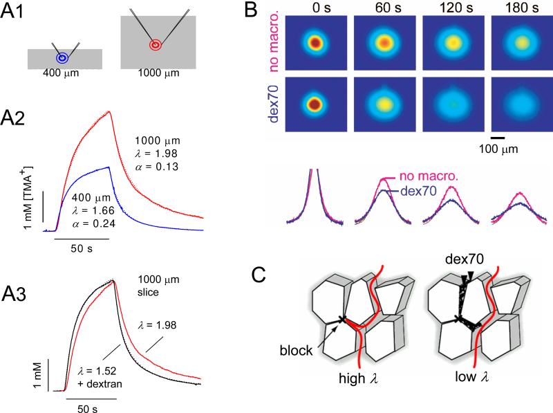
Fig. 11.
Combined use of RTI-TMA and IOI methods to investigate dead-space microdomains. A1-A3. RTI-TMA studies. A1. Schematic of independent iontophoresis microelectrode and ISM placement in a normal 400 μm slice and a 1000 μm ‘thick’ slice. The thick slice represents a model of ischemic tissue. A2. TMA+ diffusion curves in brain slices. TMA+ pulse applied for 50 s (horizontal bar) and detected by a TMA+-ISM 100 μm away. Diffusion curves are superimposed with theoretical curve (Equations 13 and 15). Representative recordings from 400 (blue curve) and 1000 μm neocortical ‘thick’ slices (red curve). As expected, the tortuosity is higher and volume fraction is lower in the ischemic slice. A3. Representative recording from 1000 μm slice incubated with 70 kDa dextran in the bath (black curve); this brings about a paradoxical fall in tortuosity. For comparison, the diffusion curve from 1000 μm slices incubated without dextran (red curve) shown in A2 is superimposed (Modified from Ref. 148). B. Use of IOI to study effect of added background molecules (70 kDa dextran without fluorescent label) on diffusion of 3 kDa dextran fluorescent molecules (fdex3). Top image row: Images of fdex3 taken immediately after the pressure injection (labeled as 0 s) and at 60, 120, and 180 s later in neocortical thick slices. The intensity shown in pseudo color (red highest, blue lowest) represents the concentration of the fdex3 in the tissue. The images in the upper row were taken in the absence of background macromolecules (no macro.). The images in the lower row were in the presence of non-fluorescent dex70. The intensity profiles of data (bottom), obtained along the horizontal line running through the center of the image. In the presence of dex70, the image intensity dissipated faster and therefore the collapse of the curve (blue) is more pronounced. Tortuosities were 3.66 and 2.37 in the absence and presence of dex70, respectively. (Modified from Ref. 147). C. It is hypothesized that during ischemia and other pathological conditions, cellular elements expand their volume as water moves from the extracellular to the cellular compartment and blockages are formed in some interstitial planes. Diffusing molecules that enter these pocket-like regions are delayed and tortuosity increases (left schematic). When background macromolecules, such as dex70, are added to this tissue, they become trapped in the dead-spaces. By excluding the dead-space volume, dex70 prevents marker molecules from being delayed there and tortuosity decreases (right schematic). (Modified from Ref. 147).

