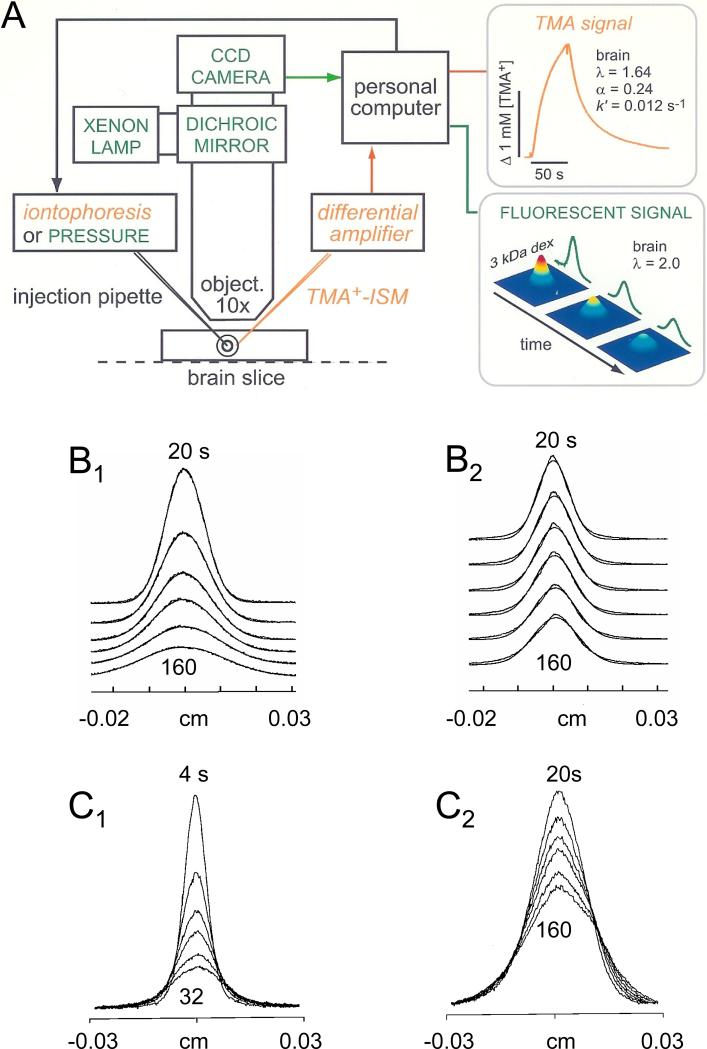
Fig. 5.
A. Setup for integrative optical imaging (IOI) method and RTI-TMA in brain slices. A brain slice or dilute agarose (or agar) gel is placed into a chamber on a stage of a compound microscope equipped with epifluorescent optics. For IOI, a fluorescent molecule is pressure injected from a micropipette and a time series of images captured using a charge-couple device (CCD) camera. The images are processed with a PC to fit Equation 20 to the intensity profiles measured along selected image axes. Diffusion coefficients, D and D*, are extracted in agarose gel and brain slice, respectively. As shown in this figure, RTI-TMA measurements can also be made in this setup with the advantage that the iontophoresis microelectrode and the ISM can be positioned independently under visual control. Both IOI and RTI measurements can be performed simultaneously. (Modified from Ref. 149). B1-B2. Diffusion profiles for 70 kDa fluorescent dextran. B1. Profiles measured at 20, 40, 60, 80, 120 and 160 s after pressure injection in agarose gel as a function of distance in object space (slice). Profiles have characteristic shape of a Gaussian curve. B2. Similar profiles measured in slices of rat cortex. Note that the profiles collapse much more slowly in brain than in agarose as a consequence of the tortuosity in the slice (λ = 2.25 for 70 kDa dextran). (Modified from Ref. 265). C1-C2. Diffusion profiles for 66 kDa fluorescent bovine serum albumin (BSA). C1. Profiles measured at 4, 8, 12, 16, 24 and 32 s after pressure injection in agarose gel as a function of distance in object space (slice). C2. Similar profiles measured in slices of rat cortex. Profiles measured at 20, 40, 60, 80, 120 and 160 s after pressure injection; again the profiles collapse much more slowly in brain than in agarose (λ = 2.26 for 66 kDa BSA). (Modified from Ref. 379).

