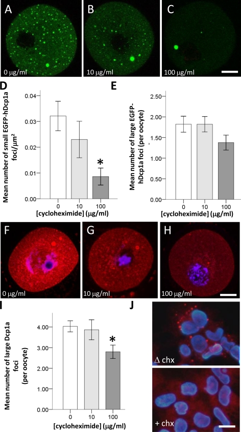
Figure 5.
Only small EGFP-hDcp1a foci are sensitive to cycloheximide. (A–C) Confocal fluorescence images showing GV oocytes microinjected with EGFP-hDcp1a and treated with either 0, 10, or 100 μg/ml cycloheximide (CHX). (A) A nontreated GV oocyte showing EGFP-hDcp1a small and large foci; (B) a GV oocyte treated with 10 μg/ml CHX still showing EGFP-hDcp1a foci; (C) a GV oocyte treated with 100 μg/ml CHX. The small foci have disappeared but the large foci are still present. (D) Graph showing the mean number of small EGFP-hDcp1a foci per μm2 for different doses of CHX; the effect of 10 μg/ml CHX (n = 56) compared with 0 μg/ml CHX (n = 45) is not significant (p = 0.45) but with 100 μg/ml CHX (n = 53) the reduction is significant (p = 1 × 10⁻³). (E) Graph showing the mean number of large EGFP-hDcp1a foci per oocyte for different doses of CHX; the effect of CHX was not significant at 10 μg/ml (p = 0.998) or at 100 μg/ml (p = 0.097). (F–H) Confocal fluorescence images of GV oocytes immunostained for Dcp1a and treated with 0 μg/ml (n = 45), 10 μg/ml (n = 50), and 100 μg/ml CHX (n = 53); large foci are present for all doses. (I) Graph showing the mean number of large Dcp1a foci per oocyte for different doses of CHX: 0 μg/ml (n = 125), 10 μg/ml (n = 30), and 100 μg/ml (n = 153), respectively; the effect of 10 μg/ml CHX was not significant (p = 0.78) but was more significant for 100 μg/ml CHX (p = 5 × 10⁻³). (J) Immunocytochemistry for Dcp1a protein in HEK-293 cells treated with (+CHX) and without (ΔCHX) cycloheximide. Scale bars, (A–C and F–H) 20 μm.

