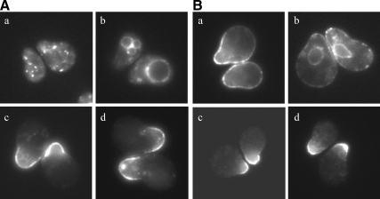
Figure 6.
The exomer-dependent IXTPK sorting signal functions in a restricted context. (A) GFP localization of (a) full-length Kex2p-GFP (percentage of cells with plasma membrane GFP: 2 ± 2), (b) C-terminally deleted Kex2p-GFP (24 ± 5), and (c) Kex2p-Fus1p-GFP (89 ± 7) in wild-type cells and (d) Kex2p-Fus1p-GFP (76 ± 13) in chs5Δ cells. All constructs contained the Fus1p transmembrane domain and were under control of the FUS1 promoter. (B) GFP localization of (a) full-length Mid2p-GFP (88 ± 9), (b) C-terminally deleted Mid2p-GFP (ND), and (c) Mid2p-Fus1p-GFP (58 ± 0) in wild-type cells and (d) Mid2p-Fus1p-GFP (60 ± 10) in chs5Δ cells. All constructs contained the Mid2p transmembrane domain and were under control of the FUS1 promoter. All cells in A and B were also mutant for endogenous FUS1 and BAR1. Cells were treated with α-factor for 90 min and then visualized by fluorescence microscopy. Numbers in parentheses are the percentage of shmooing cells with GFP localized to the plasma membrane ± the 95% confidence interval. Numbers are the average of at least two independent experiments where 50–200 cells were scored for each experiment. Plasma membrane localization was not determined for the C-terminally deleted Mid2p-GFP due to its ER localization; cortical ER localization was difficult to distinguish from plasma membrane localization.

