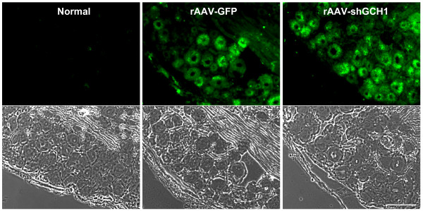
Figure 3.
Efficient gene delivery into DRG through sciatic nerve injection. The left sciatic nerves of rats were injected with rAAV-GFP or rAAV-shGCH1. At 14 days after injection, rats were sacrificed and L5 DRGs removed following perfusion. DRG tissue sections were prepared at a thickness of 10 μm. Fluorescent green signals were identified as GFP expression in both virus-treated groups under a fluorescent microscope. Scale bar = 100 μm.

