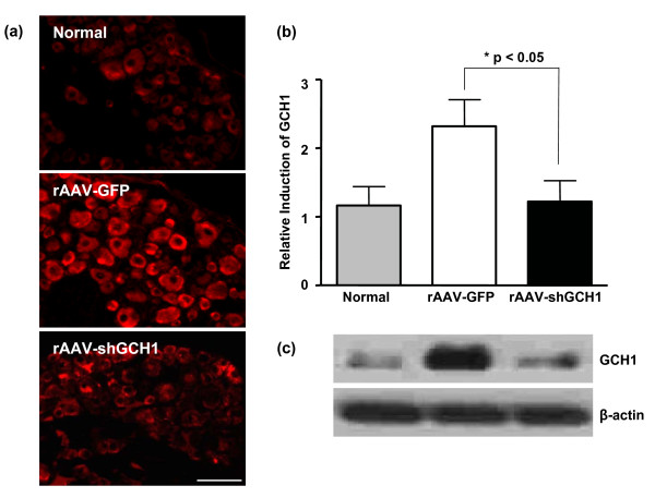
Figure 6.
Downregulation of the rGCH1 level by rAAV-shGCH1 in ipsilateral DRG. Immunohistochemistry (a, b) and western-blotting analysis (c) using GCH1-specific primary antibody allowed the visualization of rGCH1 expression 14 days after virus administration. Secondary antibody conjugated with Alexa Fluor-555 was used. The relative intensity of GCH1 expression was quantified by counting immune-positive cells following immunohistochemistry (b). Data are presented as means ± SEM (n = 8 per each groups). *p < 0.05, Scale bar = 100 μm.

