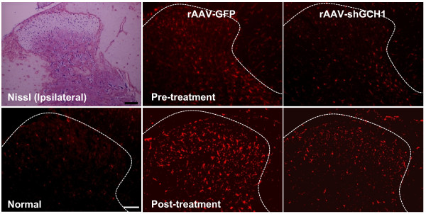
Figure 8.
Inhibition of microglial activation via rAAV-shGCH1 in the spinal cord dorsal horn. Nissl-staining was carried out in the frozen sections of spinal cords. Microglia in the spinal cord dorsal horn were visualized with the Iba-1 primary antibody on day 14 after either pain surgery for pre-treatment and virus administration for post-treatment. Immunohistochemistry data reveal that the number of Iba-1 positive cells increased in the rAAV-GFP pain group only. Scale bar = 100 μm.

