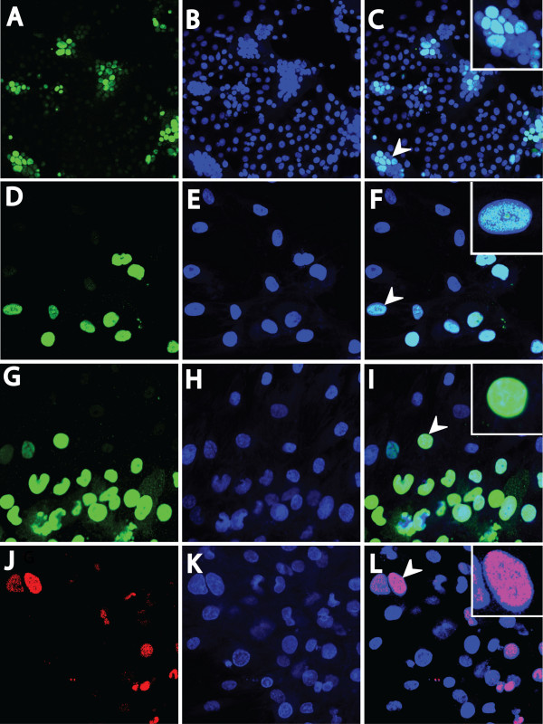
Figure 6.
The RV2 ORF59 proteins are highly expressed during RV2 rhadinovirus infections of RPFF and Vero cells and localize to the nucleus. Subconfluent cell cultures were infected with either RRV or MneRV2, and ORF59 expression was detected by confocal immunofluorescence microscopy using the anti-RV2 ORF59 antiserum. Nuclear DNA was visualized with Topro-3 stain. ORF59 immunofluorescence (A, D, G, and J), Topro-3 nuclear fluorescence (B, E, H and K), and an overlay of ORF59 and Topro-3 fluorescence (C, F, I, and L) are shown. (A-C) Vero cells infected with RRV and reacted with the rabbit 425 anti-RV2-ORF59 antiserum (10× magnification). (D-F) RPFF cells infected with RRV and reacted with the 425 antiserum (40× magnification). (G-I) RPFF cells infected with MneRV2 and reacted with 425 antiserum (40× magnification). (J-L) RPFF cells infected with MneRV2 and reacted with mouse anti-HHV8 ORF59 monoclonal antibody (40× magnification). Arrows indicate the cell/s shown in the inserts with evidence of syncytia/aggregation (C), and co-localization of RV2 ORF59 and nuclear DNA (F, I and L).

