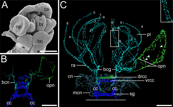
Figure 6.
Development of operculum and radiole innervationin Spirorbis cf. spirorbis. Scanning electron micrograph (A) and 3D reconstructions of serotonin immunoreactivity in corresponding body regions (B, C). Scale bars: 40 μm. Apical is to the top. (A) Dorsal view with the collar (co) enclosing the rudiments of the branchial crown (br) and the operculum (op). (B) Dorsal view, recently settled larva. Neurons of the branchial crown (bcn; in turquoise), the operculum (opn; in green), and the circumesophageal connectives (cc; in blue) are visible. Note that development of the neuronal innervation of the operculum is ahead of neurogenesis of the radioli. (C) Ventral view, settled juvenile. The neuronal innervation of the branchial crown (turquoise) shows no connection to the one of the operculum (green). The latter comprises a loop-like opercular nerve (opn) and neurons that irregularly interconnect its two branches (arrowheads). The filaments of the branchial crown, radioli (ra), and pinnulae (pi) are each innervated by three nerves (inset) that branch off from the two branchial crown ganglia (bcg). A clear designation of radioli and pinnulae is partly hindered by their similar appearance at this developmental stage. Therefore, the distal tips of the branchial filaments are consecutively numbered (1-8), indicating their symmetrical order. Note the concentration of the central nervous system (blue) with shortened circumesophageal connectives (cc) and newly formed subesophageal ganglia (sg). The neuronal network of the collar (cn) with the two main collar neurons (mcn) is shown in semi-transparent blue-grey.

