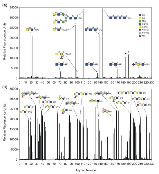
Figure 2. Sugar-binding specificities of CGLs.
(a) Binding of CGL3 to carbohydrates of the Consortium of Functional Glycomics (CFG) glycan array. Results shown are averages of triplicate measurements of fluorescence intensity. Error bars indicate the standard deviations of the mean. Glycans marked with an asterisk were not considered in the analysis due to heterogeneity. (b) CGL2 binding to the CFG glycan array. Raw data as well as the entire list of glycans with their respective spacers (SP) can be found on the CFG homepage (http://functionalglycomics.org) and in supplementary Table 1. S stands for sulphate.

