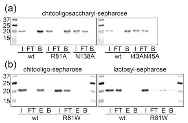
Figure 6. Solid-phase binding assay using WT and mutated CGL3.
Coomassie-stained SDS-PAGE gels of proteins tested with chitooligosaccharyl- and lactosyl sepharose are shown. Abbreviations in the figure stand for the following terms: I (input), FT (flow through), E (elution) bound fraction specifically released using the respective sugars in solution, B (beads) residual protein on column detached by boiling the sepharose beads in protein sample buffer. Equivalent amounts of each fraction were loaded on the gel. (a) binding of WT and R81A, N138A and I43AN45A mutant forms of CGL3 to chitooligosaccharyl-sepharose. (b) binding of WT and R81W mutant form of CGL3 to chitooligosaccharyl- and lactosyl-sepharose.

