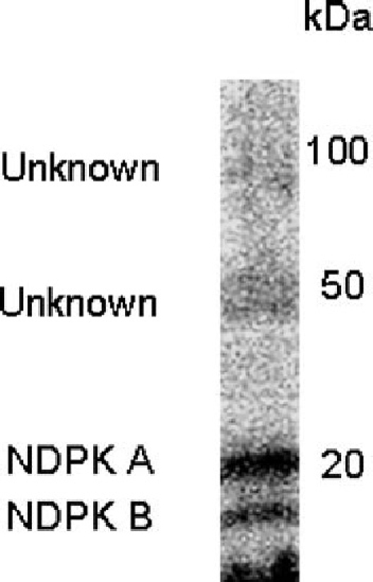
Fig. 2.

“In-gel“ autophosphorylation of proteins in membrane of ovine airway epithelium. Apical membrane proteins from sheep tracheal epithelia were prepared by sucrose gradient technique and separated by SDS-PAGE (12.5%). Following re-naturation “in-gel“ (Muimo et al., 1998), proteins were autophosphorylated by incubating the gel with 9.25 MBq γ[32P]-ATP (16 nm, specific activity 6000 Ci/mmol) in 50 ml of 25 mM MOPS pH 7.9, 10 mM MnCl2 and 0.05% Triton X-100. Labelled proteins were then detected by electronic autoradiography. Result is representative of at least three separate experiments. This figure has been adapted and reproduced with permission from Am J Resp Cell Mol Biol.
