Fig. 1.
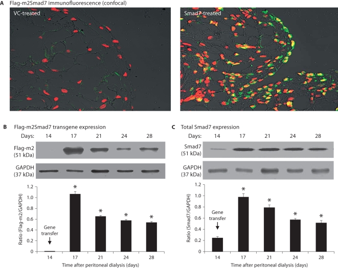
Ultrasound-microbubble-mediated Smad7 transfection rate and transgene expression in fibrotic peritoneal tissues. A Dox-regulated flag-m2-Smad7 pTRE plasmid and a Tet-on plasmid (1:1) were mixed with Sonovue at a ratio of 1:1 (vol:vol) and transfected into the diseased peritoneum on day 14 after PD by the ultrasound-microbubble-mediated system. Smad7 transgene expression was detected by confocal microscope and Western blot with the anti-Flag-M2 antibody as described in the Methods. A Confocal microscopy shows a strong Flag-m2Smad7 expression (green) in mesothelial cells on the peritoneal surface(s) and also in cells in submesothelial areas on day 3 after gene transfer (day 17 of the PD model), accounting for up to 80% of peritoneal cells, but this is negative in the control vector treatment. ×400. B Western blot analysis demonstrates Flag-m2Smad7 transgene expression over the 2-week period after gene transfer (days 14–28 of the PD model). C Western blot analysis of total peritoneal Smad7 expression over days 14–28 after gene transfer on day 14 of the PD model. Note that there is a minor Smad7 expression in the diseased peritoneal tissues on day 14 (C), which is largely increased after gene transfer of Smad7. Data represent the mean ± SD for groups of 6 rats. ∗ p <0.01 vs. day 14 group. Nuclei were counterstained with DAPI (red).

