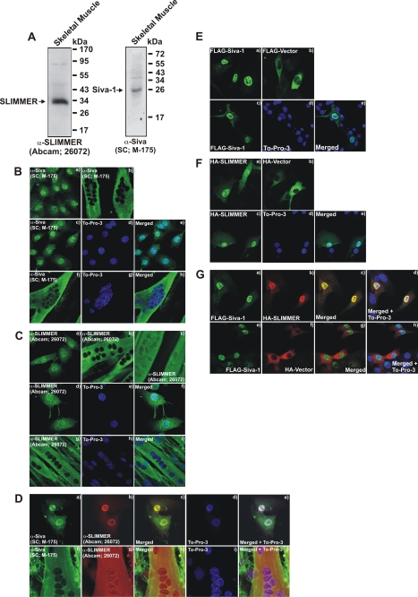
FIGURE 4.
SLIMMER and Siva-1 co-localize in C2C12 skeletal myoblasts and myotubes. A, Siva and SLIMMER are expressed in murine skeletal muscle lysates. The gastrocnemius muscle was dissected from wild type mice, the tissue was homogenized, and protein was extracted using 1% Nonidet P-40 lysis buffer. 50 μg of Nonidet P-40-soluble lysates was separated on 12.5% SDS-PAGE and immunoblotted with a goat polyclonal SLIMMER antibody (Abcam 26072) or rabbit polyclonal Siva antibody (Santa Cruz Biotechnology M-175) as indicated. B and C, localization of endogenous Siva and SLIMMER in undifferentiated C2C12 myoblasts or differentiated myotubes. Myoblasts were plated onto fibronectin-coated coverslips, cultured for 24 h, fixed and stained with either a rabbit polyclonal Siva-specific antibody (B-a) or a goat polyclonal SLIMMER-specific antibody (C-a), and co-stained with To-Pro-3 iodide to detect nuclei (B, c–e, Siva) (C, d–f, SLIMMER) as indicated. Alternatively, myoblasts were cultured for a further 24 h until confluent and switched to low serum media to induce differentiation to multinucleated myotubes. Myotubes were also fixed and stained with either a rabbit polyclonal Siva-specific antibody (B-b) or a goat polyclonal SLIMMER-specific antibody (C, b and c) and co-stained with To-Pro-3 iodide to detect nuclei (B, f–h, Siva; C, g–i, SLIMMER) as indicated. D, endogenous Siva and SLIMMER co-localize in myoblasts and myotubes. Myoblasts (D, a–e) and myotubes (D, f–j) were prepared as described in B and C and co-stained with rabbit polyclonal Siva (D, a and f), goat polyclonal SLIMMER (D, b and g) antibodies, and the nuclear stain To-Pro-3 iodide (D, d and i) as indicated. E–G, C2C12 myoblasts were transiently transfected with FLAG-Siva-1 (E, a and c–e), FLAG-vector (E-b), HA-SLIMMER (F, a and c–e), or HA-vector (F-b) as indicated and stained with anti-FLAG (E) or anti-HA (F) antibodies, respectively. A subset of cells were also co-stained with To-Pro-3 iodide to detect nuclei (E, c–e, and F, c–e). G, FLAG-Siva-1 co-localizes with HA-SLIMMER in myoblasts. C2C12 myoblasts were co-transfected with FLAG-Siva-1 and either HA-SLIMMER (G, a–d) or HA-vector (G, e–h). Cells were co-stained with anti-FLAG and anti-HA antibodies and To-Pro-3 to detect nuclei. All cells were viewed using laser-scanning confocal microscopy.

