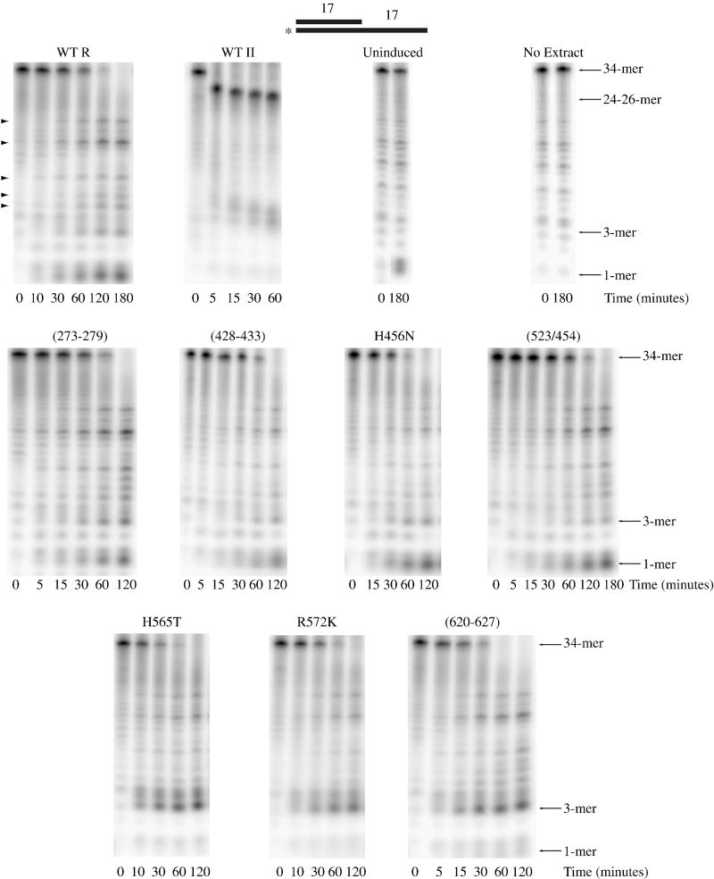
Figure 2. Activity of Nuclease Domain Channel Mutant Extracts on Structured RNA.
Assays were carried out as described under “Materials and Methods” with 10 μM ds17-A17 substrate and 2.5 μg wild-type RNase R, 0.5 μg wild-type RNase II, 2.5 μg uninduced, 2.5 μg RNase R (273-279), 2.5 μg RNase R (428-433), 5 μg RNase R H456N, 2.5 μg RNase R (523/545), 10 μg RNase R H565T, 2.5 μg RNase R R572K and 2.5 μg RNase R (620-627) cell extract. Aliquots were taken at the indicated times and analyzed by denaturing PAGE. A schematic representation of the substrate is shown at the top of the figure with the position of the 32P label denoted by an asterisk. Non-specific product bands that do not appear to originate from RNase R activity are indicated on the left of the wild-type RNase R panel by arrowheads.

