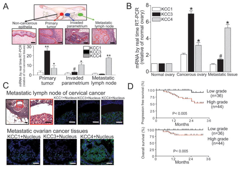
Figure 1. KCC4 expression is associated with cancer metastasis and clinical outcome.

(A) & (B) KCC4 is abundant in metastatic tumor tissues. Laser microdissection with microscopic observation was utilized to sample the targeted tissues precisely (dashed line). The KCC mRNA levels were normalized against GAPDH. The KCC expression levels in normal squamous epithelia and normal ovaries were used as the control for the cases of cervical cancer and ovarian cancer, respectively. Each column represents mean ± S.E.M. (n=8 for cervical cancer; n=6 for ovarian cancer). #P<0.05; *P<0.01; **P<0.001 by paired t test. Scale bar, 10μm. (C) The expression pattern of KCC family in metastatic pelvic lymph nodes of cervical cancer (representative images of 6 different cases; scale bar, 5μm) and metastatic tissues of ovarian cancer (representative images of 6 different cases; scale bar, 10μm). (D) The association between KCC4 expression and clinical outcome at early stage cervical cancer. Cervical cancer patients were grouped by KCC4 grading and the survival data analyzed accordingly.
