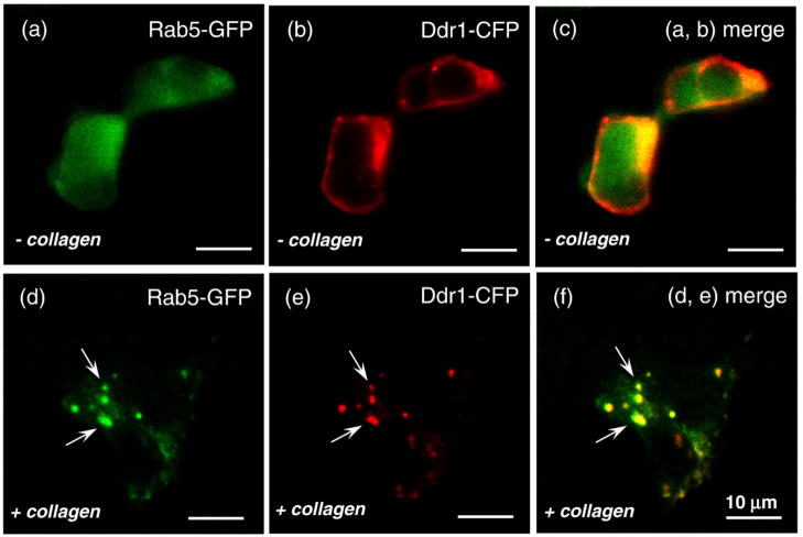
Fig. 10.
DDR1 aggregates colocalize with Rab5a. HEK293 cells were transiently transfected with both DDR1–CFP and Rab5a-GFP and stimulated with collagen type I for 15 min (as indicated). Confocal images of nonstimulated cells are shown on the upper row (a–c), and stimulated cells are shown on the lower row (d–f). CFP, red; GFP, green; overlap of the two, yellow. (f) The aggregated DDR1 (indicated by white arrows) in the stimulated cells overlaps with Rab5a. All scale bars represent 10 μm.

