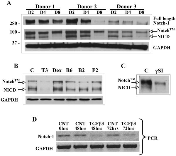
Figure 1. Reduced Notch-1 activity during chondrogenic differentiation is mediated by TGFβ exposure.
Cell lysates were prepared from pellet cultured human MSC at days 2, 4 or 8 from three donors (panel A). Western blots showed a reduction in NICD and full length Notch-1 protein levels were consistently seen by 8 days of pellet culture. To test whether the prochondrogenic factors added during chondrogenesis induced reduction in NICD levels, hMSC were exposed to TGFβ3 (T3; 10 ng/ml), Dexamethasone (Dex; 10−8 M), BMP6 (B6; 500 ng/ml), BMP2 (B2; 500 ng/ml) and FGF2 (F2; 50 ng/ml) in monolayer culture for 72 hours. Western blot of NICD indicated that TGFβ3 was responsible for the reduction in Notch-1 protein levels (panel B). Notch™ = Transmembrane portion of Notch-1 plus the uncleaved NICD fragment. In monolayer cultured hMSC addition the gamma secreatase inhibitor (γSI) L685,458 at a concentration of 5 μM for 24 hours prevented cleavage of NICD, indicating that the bTAN20 antibody detects both Notch™ and cleaved Notch-1 intracellular domain (NICD) proteins (panel C). RT-PCR (panel D) showing expression levels of Notch-1 mRNA in monolayer cultured hMSC incubated with TGFβ3 (10 ng/ml) for 48 and 72 hrs. Reduction in Notch-1 protein levels is regulated by TGFβ3 at the transcription level.

