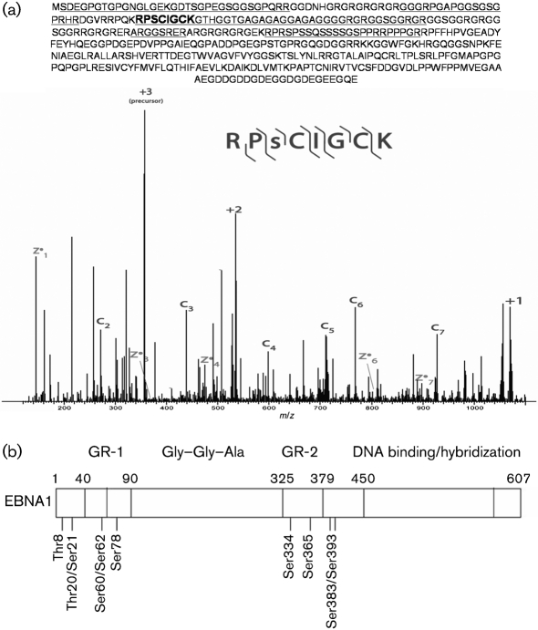
Fig. 1.
Identification of EBNA1 phosphosites. (a) Representative spectrum of a phosphorylated EBNA1 peptide. The location of this residue within the protein is shown in bold and underlined. C ions correspond to N-terminal fragments of the peptide recovered, whilst Z ions correspond to C-terminal fragments. The total peptides that were recovered in all of the ETD-MS/MS runs are underlined. The +1, +2 and +3 m/z peaks correspond to the precursor molecule that has undergone electron transfer, but has not dissociated. (b) Schematic of the EBNA1 protein showing the location of the phosphosites within the domain structure. The NLS is located between aa 379 and 386.

