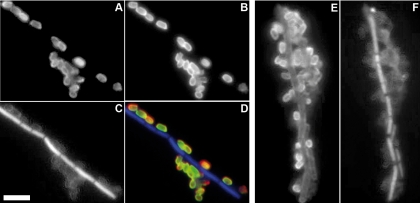
FIG. 4.
Immunofluorescence staining of B. anthracis using polyclonal antibodies against anthrose and spores. Inactivated B. anthracis spores were stained with polyclonal anthrose antibody pc115 (A) and were visualized with a goat anti-rabbit Alexa 594-labeled secondary antibody. Colocalization with the BclA protein (B) was shown by using monoclonal BclA antibody EG4-4, detected with goat anti-mouse Alexa 488-labeled antibody. The overlapping of the staining of pc115 and EG4-4 appears yellow and is shown as a merged image (D). The H64-BA IgY polyclonal spore antibody (E) was visualized by using goat antichicken Alexa 488-labeled detection antibody. The presence of spores was observed by phase-contrast microscopy (data not shown), and cell nuclei were stained with DAPI (C and F). Note the staining only of spores and not of vegetative cells in the case of the anthrose and BclA antibodies. Magnification, ×100, oil immersion. Bar, 10 μm.

