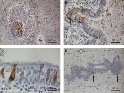
FIG. 3.
Immunohistochemistry of lung and uterus tissue from animal R3. The sections were labeled with a mouse monoclonal antibody to CvHV2 envelope gC, and positive labeling was visualized by a horseradish peroxidase-DAB method. Hematoxylin was used as a counterstain. (A) Cranioventral lung with a necrotic bronchiole in the area with bronchopneumonia. Positive immunolabeling for CvHV2 is seen as a brown stain in the tissues. BL, bronchiole lumen; BV, blood vessel; Alv, alveole filled with exudate. Magnification, ×200. (B) Bronchi in caudodorsal lung. Degenerated, CvHV2-positive cells (brown-stained cells, arrows) are seen in the respiratory epithelium, and positively stained cell debris (brown stain) is seen in the bronchial lumen (L). Magnification, ×400. (C) Highly magnified view of respiratory epithelium showing CvHV2-positive goblet cells (brown-stained cells). Magnification, ×1,000. (D) CvHV2-positive uterine cells (brown cells, arrows) in an endometrial gland. Magnification, ×400.

