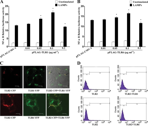
FIG. 4.
TLR1 and TLR6 enhanced TLR2-mediated NF-κB activation. (A) HEK293T cells were transiently cotransfected with the indicated concentrations of pFLAG-TLR1, 0.1 μg ml−1 pFLAG-TLR2, 0.1 μg ml−1 pNF-κB-luc, and 0.01 μg ml−1 pRL-TK. After 24 h of incubation, the cells were stimulated for 8 h with 2.0 μg ml−1 LAMPs. The cells then were lysed and assayed for luciferase reporter activity. All values represent the means and standard deviations from three assays. P < 0.05 (*) was considered significant. (B) HEK293T cells were transiently cotransfected with the indicated concentrations of pFLAG-TLR6, 0.1 μg ml−1 pFLAG-TLR2, 0.1 μg ml−1 pNF-κB-luc, and 0.01 μg ml−1 pRL-TK. After 24 h of incubation, the cells were stimulated for 8 h with 2.0 μg ml−1 LAMPs. The cells then were lysed and assayed for luciferase reporter activity. All values represent the means and standard deviations from three assays. P < 0.05 (*) was considered significant. (C) HEK293T cells were transiently cotransfected with TLR2-CFP, and TLR1-YFP or TLR6-YFP was grown on glass-bottomed tissue culture dishes. After 24 h of incubation, the cells were stimulated for 8 h with 2.0 μg ml−1 LAMPs. The living cells then were analyzed by confocal microscopy as described in the text. To the left is the distribution of TLR2; in the center is the localization of TLR1 or TLR6; to the right is the colocalization of TLR2 with TLR1 or TLR6 (black arrows or white arrows). Representative confocal sections of cells are shown. (D) HEK293T cells were transiently cotransfected with the indicated constructs. After 24 h of incubation, the cells were stimulated for 8 h with 2.0 μg ml−1 LAMPs. The cells then were incubated with anti-TLR2 MAb and FITC-labeled secondary Ab. The cell surface expression of TLR2 was analyzed by flow cytometry. Representative confocal sections of cells are shown.

