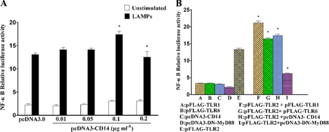
FIG. 5.
CD14 enhanced TLR2-mediated NF-κB activation. (A) HEK293T cells were transiently cotransfected with the indicated concentrations of pcDNA3-CD14, 0.1 μg ml−1 pFLAG-TLR2, 0.1 μg ml−1 pNF-κB-luc, and 0.01 μg ml−1 pRL-TK. After 24 h of incubation, the cells were stimulated for 8 h with 2.0 μg ml−1 LAMPs. The cells then were lysed and assayed for luciferase reporter activity. All values represent the means and standard deviations from three assays. P < 0.05 (*) was considered significant. (B) HEK293T cells were transiently cotransfected with the indicated plasmids (the concentration of each plasmid was 0.1 μg ml−1). After 24 h of incubation, the cells were stimulated for 8 h with 2.0 μg ml−1 LAMPs. The cells then were lysed and assayed for luciferase reporter activity. All values represent the means and standard deviations from three assays.

