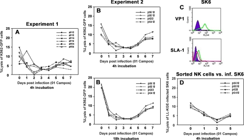
FIG. 2.
Cytotoxic activity of porcine peripheral blood NK cells. PBMC were isolated from noninfected pigs or pigs infected with O1 Campos strain of FMDV, and NK cell lytic assays against K562-GFP cells in a 4-h assay were performed daily until day 7. (A and B) Two different sets of animal groups were analyzed in two different experiments out a total of three separate experiments. (C) SK6 cells infected (inf.) with LL-KGE, VP1, and SLA-I were assessed by flow cytometry. The shaded area of the upper panel is VP1 staining in noninfected cells, and the green line is VP1 staining in infected cells; the shaded area of the lower panel is the isotype monoclonal antibody control, the green line is the SLA-I staining of noninfected cells, and the red line is the SLA-I staining of virus-infected cells. (D) CD2+/CD8+/CD3− cells were purified from PBMC on days 0, 1, 3, and 5, and the killing assay was performed on SK6 cells previously infected with LL-KGE virus. Data represent a single experiment from a total of two separate determinations. Data represent a single experiment from a total of two. Percent lysis at an E:T ratio of 50:1 is shown. Numbers in the graph legend denote individual animal identification tags.

