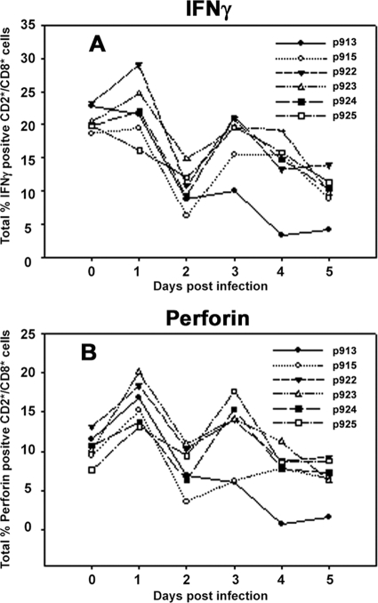
FIG. 5.
IFN-γ and perforin profile in infected animals. PBMC were isolated on 5 consecutive days from infected animals and stained intracellularly for IFN-γ and perforin as described in Materials and Methods. CD3+ cells were gated out, and another gate was created on either IFN-γ- or perforin-positive cells and then applied to CD3−/CD2+/CD8+ cells to give the percentage of CD2+/CD8+ positive cells for either IFN-γ (A) or perforin (B). Data represent a single example from five separate experiments. Day 0 represents data derived from noninfected animals and serves as a point of reference. Numbers in the graph legend denote individual animal identification tags.

