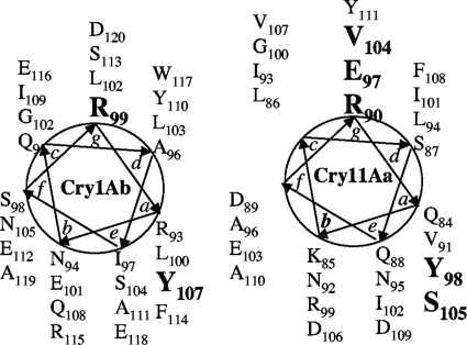
FIG. 1.
Schematic representation of the coiled-coil structures of the α-3 helices of Cry1Ab and Cry11Aa toxins. The positions of residues a, b, c, d, e, f, and g of the heptads are presented. The mutated residues in both toxins that affected oligomerization and toxicity are shown in boldface type (reference 4 and this work).

