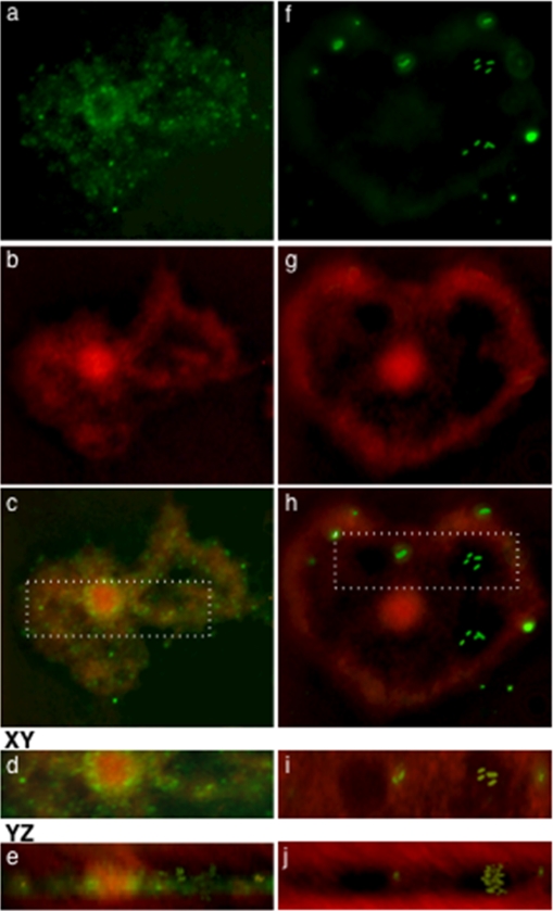
FIG. 4.
Immunofluorescence z-stack projections of A. castellanii trophozoites infected with LVS expressing GFP for 2 h (a, b, and c) and NOV stained with an anti-F. tularensis antibody (green) (f, g, and h) after 2 h and 30 min, respectively. At 2 h postinfection, the majority of LVS bacteria (a) colocalize with LysoTracker red (c), which appears diffuse within the trophozoite (b). The majority of NOV bacteria (f) do not colocalize with LysoTracker red (h), which appears localized (g). Enlarged cross sections of panels c and h represent trophozoites infected with LVS (d) and NOV (i). These were rotated by 90° using Volocity software to confirm the presence of the bacteria intracellularly (e and j).

