FIG. 5.
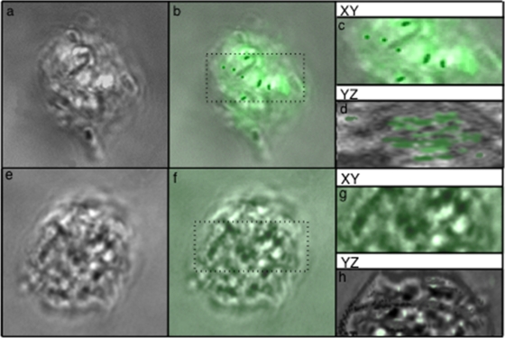
Nomarski DC z-stack projections of uninfected A. castellanii cysts (e and f) and cysts infected with NOV (a and b) and stained with an anti-NOV antibody (b and f). Enlarged cross sections of panels b and f (c and g) were rotated by 90° using Volocity software to confirm the presence (d) or absence (h) of intracellular bacteria.

