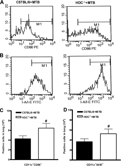
FIG. 6.
HDC−/− mice demonstrated enhanced activation and frequency of APCs in the lungs upon M. tuberculosis infection. Lung tissue was removed at 28 days after M. tuberculosis H37Rv infection (102 CFU intranasally), and lung cells from M. tuberculosis-infected C57BL/6 (black bars in panels C and D) or HDC−/− (white bars in panels C and D) mice were analyzed by flow cytometry. Representative histograms of the expression of CD86 (A) and MHC class II (B) in a gated CD11c-positive cell population are shown. The presence of CD11c+ CD86+ or CD11c+ IA IE cells is expressed as absolute number of positive cells (C and D). The means ± SEM from at least five mice per group in one independent experiment repeated twice are shown. Numeral signs represent a significant difference (P < 0.05) relative to M. tuberculosis-infected C57BL/6 mice. Statistical variations were analyzed by Student's t test.

