FIG. 7.
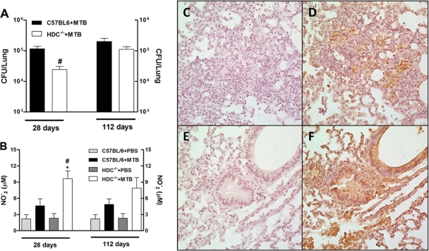
Accelerated clearance associated with profound iNOS and NO expression in the lungs of HDC−/− mice was observed after M. tuberculosis infection. (A) The mycobacterial burden was enumerated by counting CFU at 28 and 112 days after M. tuberculosis H37Rv infection (102 CFU intranasally). (B) Nitrite production was quantified by Griess reaction in the lung homogenates recovered at the same time points. (C to F) Expression of iNOS in lung tissue of infected C57BL/6 (D) or HDC−/− (F) mice was analyzed by immunostaining on day 28 after infection. Brown staining shows iNOS-positive cells. iNOS staining without primary antibody of infected C57BL/6 (C) or HDC−/− (E) mice was used as a background control. Results are expressed as means ± SEM from at least five animals per group in one independent experiment repeated twice. In panels A and B, significant differences (P < 0.05) relative to uninfected (*) or M. tuberculosis-infected (#) C57BL/6 mice are shown. PBS, phosphate-buffered saline.

