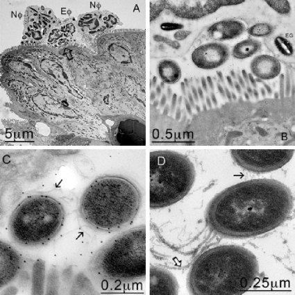
FIG. 5.
PMNs engulfing C. difficile B1 at the mucosal membrane. (A) TEM image showing neutrophils (Nφ) and eosinophils (Eφ) at the mucosal surface that appear to have engulfed several bacteria. (B) Higher magnification of the area indicated by the arrow in panel A, confirming that C. difficile was trapped against the mucosal membrane by the host cells. (C) Higher magnification of panel B. The bacteria were confirmed to be C. difficile by immunogold labeling. (D) In contrast, 630 bacteria found in the crypts appear to express both flagella (open arrow) and pili (solid arrow).

