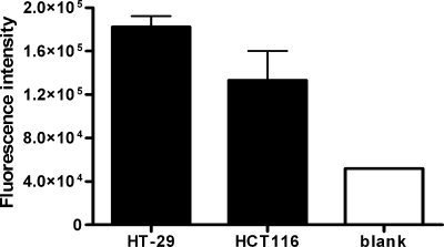
FIG. 3.
Binding of HlpA to intact adenocarcinoma cells. In-cell Western analysis of HT-29 and HCT116 colorectal cancer cells incubated with purified HlpA-His from S. gallolyticus. Detection of HlpA-His was established by incubation with rabbit anti-HlpA antibody and with Alexa Fluor 680-labeled secondary anti-rabbit antibody. The bars represent the fluorescence intensities of HlpA-His on adenocarcinoma cells. The fluorescence intensity of HlpA was corrected for the basal fluorescence level (nonspecific fluorescence) of the indicated cell line. The blank measurement was obtained by subtracting the fluorescence intensity of a blank well incubated with DMEM from that of a well incubated with DMEM with HlpA (background measurement). Error bars indicate the standard errors from two replicate experiments.

