FIG. 2.
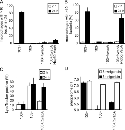
Characterization of R. equi 103+/ΔvapA virulence phenotypes. (A) Multiplication in J774E of 103+, 103−, 103+/ΔvapA, and 103+/ΔvapA complemented with vapA constitutively expressed from hsp60 promoter (103+/ΔvapA Hsp60-VapA). The percentages of infected macrophages with more than 10 bacteria are indicated. Quantitation was done microscopically, based on staining of bacterial DNA with SYTO13. The data represent means and standard deviations from three independent experiments. (B) Same as in panel A, but complementation was done using the endogenous vapA promoter and the vapA gene (103+/ΔvapA endog.VapA). (C) Qualitative determination of phagosome acidification. J774E were infected as in panel A and, after 2 or 24 h, the samples were stained with red LysoTracker and green fluorescent SYTO13. Colocalization between LysoTracker- and green-labeled bacteria was quantified by using confocal laser scanning microscopy. The percentages of phagosomes positive for LysoTracker are indicated. The data shown are the means and standard deviations of three experiments. In panels A, B, and C, at least 50 infected macrophages were analyzed in each experiment per time and sample type. (D) Quantitative determination of average phagosome pH at 3 h of infection (▪). The addition of nigericin collapses pH gradients between phagosome and cytosol and serves as a control for the calibration process in that the pH should be that of the external buffer (∼7.3) after addition (□). Nigericin was added at 3 h of infection (first pH determination), followed by a 20-min incubation period (redetermination of pH [□]).

