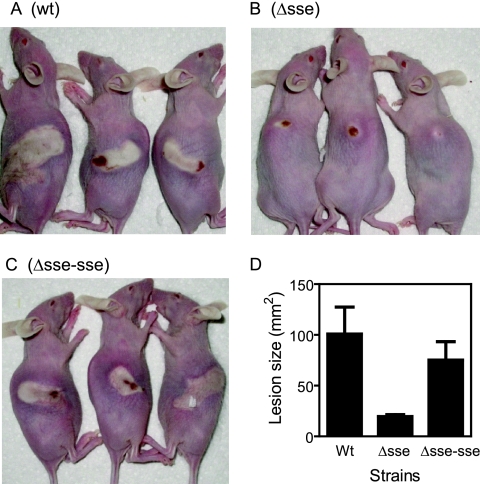
FIG. 5.
Comparison of the skin lesions resulting from subcutaneously infected WT, Δsse, and Δsse-sse strains. Immunocompetent hairless female mice (strain Crl:SKH1-hrBR) were subcutaneously infected with 50 μl of 9.8 × 10⁷ CFU of MGAS5005 (A), 9.6 × 10⁷ CFU of the Δsse strain (B), or 9.4 × 10⁷ CFU of the Δsse-sse strain (C). Panels A through C show representative infection lesions of three mice from each group on day two following infection. Panel D shows the mean size ± standard deviation for the lesions in each group at day 2.

