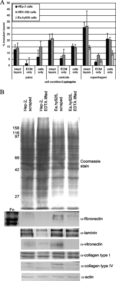
FIG. 2.
Attachment of Leptospira to cells versus the ECM. (A) Confluent cell layers were left as is (intact layers) or lifted with EDTA twice to remove all cells from the ECM. The cells were collected by centrifugation, and both the cells and the ECM were washed in medium without antibiotics to remove the EDTA and restore the divalent cations prior to the addition of 35S-labeled Leptospira bacteria at a multiplicity of infection of 10. After incubation for 1 h at 37°C, all wells were washed to remove unbound bacteria, and bound bacteria were quantified by scintillation counting. The results presented were calculated by subtracting the level of background binding to wells without cells or ECM material. The means and standard deviations of results for four replicates from representative experiments (≥4 for each cell line) are shown and are expressed as the percent inoculum bound. By using Student's two-tailed t test, comparisons of binding to lifted cells versus binding to the ECM left behind were as follows: for HEp-2 cells, P > 0.1 for L. biflexa serovar Patoc and P = 0.0002 for L. interrogans serovars Canicola and Copenhageni; for HEK293 cells, P = 0.008 for L. biflexa serovar Patoc, P > 0.06 for L. interrogans serovar Canicola, and P < 0.0001 for L. interrogans serovar Copenhageni; and for Ea.hy926 cells, only serovar Copenhageni was tested, with P < 0.0001 for cell binding versus ECM binding. (B) Immunoblots of ECM proteins associated with cells after either scraping of the cell layers into SDS gel loading buffer or lifting of the cells with EDTA prior to solubilization in gel loading buffer. As a positive control for the antifibronectin (α-fibronectin) antibody, 0.1 μg of purified fibronectin (Fn; soluble form) was loaded. The first lane of the blot probed with antivitronectin (α-vitronectin) was taken from one of the other panels after reprobing with antivitronectin, as the original blot was miscut between lanes. Molecular size markers (in kilodaltons) are shown to the left. α-laminin, α-collagen type I, α-collagen type IV, and α-actin, antilaminin, anti-collagen type I, anti-collagen type IV, and antiactin antibodies.

