FIG. 6.
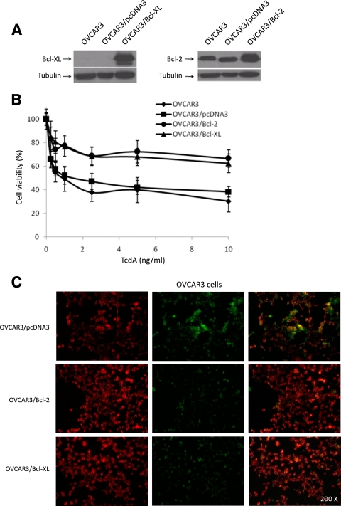
Effects of Bcl-2 and Bcl-XL overexpression on TcdA-induced cell death in OVCAR3 cells. (A) Immunoblot analysis of OVCAR3 cells stably transfected with either empty vector (OVCAR3/pcDNA3), Bcl-2 (OVCAR3/Bcl-2), or Bcl-XL (OVCAR3/Bcl-XL). (B) Effects of enforced expression of Bcl-2 and Bcl-XL on the viability of OVCAR3 cells in response to a 48-h treatment with increasing concentrations of TcdA. The data are means ± SEM of three independent experiments performed in triplicate. P < 0.001 relative to parental OVCAR3 and OVCAR3/pcDNA3. (C) OVCAR3/pcDNA3 (control), OVCAR3/Bcl-2, and OVCAR3/Bcl-XL cells were cultured for 24 h without TcdA, and the mitochondrial-membrane integrity was assessed using the MitoLight apoptosis detection kit. For treated cells, fresh culture medium containing 20 ng/ml of TcdA was added for 5 h prior to MitoLight staining. The red fluorescence (left column) represents dimeric dye that accumulated in the intact mitochondrial membrane, indicating nonapoptotic cells. The green fluorescence (middle column) represents cytoplasmic pools of monomeric lipophilic cationic dye, indicating the inability of mitochondria to concentrate the dye, and consequently shows apoptotic cells. The right column contains overlays of the left and middle columns.

