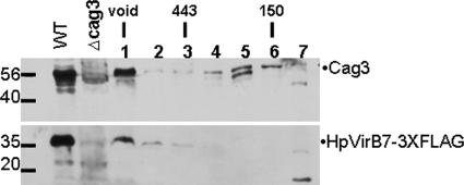
FIG. 5.
Cag3 and HpVirB7-3XFLAG fractionate as a high-molecular-mass complex. A whole-cell extract of NSH57, virB7::3XFLAG::cat (WT), was fractionated on a Superdex 200 gel filtration column, and the presence of Cag3 and HpVirB7-3XFLAG in each fraction was determined by immunoblotting with anti-Cag3 or anti-FLAG antibody. Molecular mass marker sizes in kDa are indicated. Fraction 1 is the void volume at which dextran blue eluted, the 443-kDa marker eluted in fraction 3, and the 150-kDa marker eluted in fraction 6. WT, HpvirB7::3XFLAG::cat; cag3, cag3::aphA3. Data shown are representative of two independent experiments.

