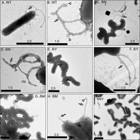
FIG. 2.
Flagellation of H. pylori fli mutants. Electron micrographs of H. pylori cells stained with phosphotungstate. In each panel, arrows mark flagella and arrowheads mark terminal bulb structures. (A) Wild-type (WT) G27; (B) detail of G27 wild-type flagella; (C) G27 ΔfliN::cat flagellated cells; (D) detail of G27 ΔfliN::cat flagella; (E) G27 ΔfliY::cat; (F) detail of G27 ΔfliY::cat mutant flagella; (G) G27 ΔfliM::cat; (H) rare G27 ΔfliM::cat flagellated cells; (I) G27 ΔfliN::cat ΔfliY::aphA3 double mutants. Bar lengths are in micrometers. The squares in panel C are staining artifacts.

