
FIG. 4.
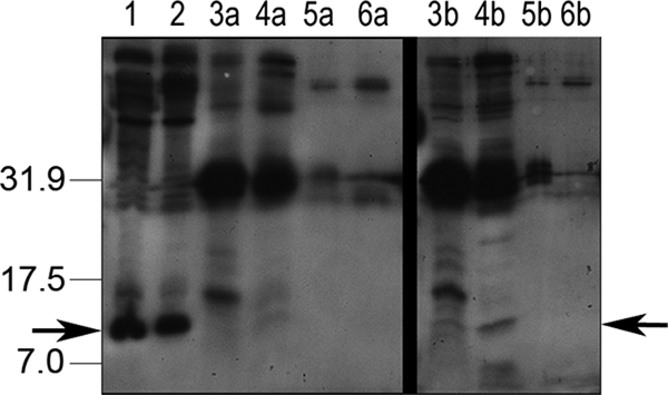
Western blot of proteins that interact with GST-FliM, using anti-CheY antisera. Specific bands consistent with CheYHP are marked with arrows. On the left are molecular mass markers in kilodaltons. Lanes: 1 and 2, presence of CheY in the starting materials (1, whole-cell extract from wild-type H. pylori; 2, whole-cell extract from H. pylori CheA); 3, GST beads plus CheY-P (wild type); 4, GST-FliM beads plus CheY-P (wild type); 5, GST beads plus unphosphorylated CheY (extract from CheA); 6, GST-FliM beads plus unphosphorylated CheY (extract from CheA). Lanes 3a to 6a were washed with 0.3 M KCl. Lanes 3b to 6b were washed with 0.4 M KCl. Higher-KCl washes resulted in the complete removal of CheY.