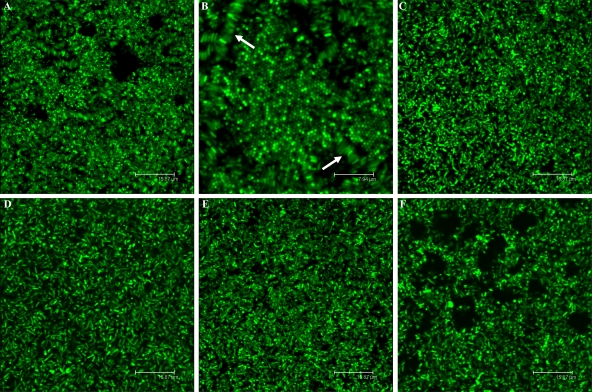
FIG. 6.
CLSM images of S. meliloti biofilms in low-phosphate MGM medium (12 dpi). The (A and B) exoY, (C) expR, (D) sinI (E) expA, and (F) expR mucR mutant strains are shown. Bars (except for panel B), 15.8 μm. Panel B shows honeycomb-like structures and lateral interactions indicated by arrows (bar, 7.9 μm).

