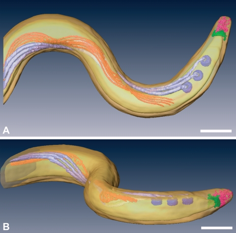
FIG. 1.
Top (A) and side (B) views of a surface-rendered model of T. pallidum, showing the outer and cytoplasmic membranes (transparent yellow), basal bodies (dark lavendar), flagellar filaments (light lavendar), cytoplasmic filaments (orange), cap (green), and cone (pink). The peptidoglycan layer was not rendered. Also see Movie S1 in the supplemental material for animation. Bars, 200 nm.

