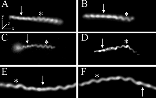
FIG. 2.
T. palladium has a flat-wave morphology. Representative micrographs show the flat-wave morphology of T. pallidum as revealed by dark-field microscopy (A and B) and by epifluorescence microscopy (C and D) following labeling of motile treponemes with FM4-64. Panels C and D show sequential images of the same treponeme; note how the leftward segment changes from helical to linear as it moves away from the focal plane. (E and F) For comparison purposes, dark-field images of a B. burgdorferi 297 cell in two different orientations are shown. Arrows and asterisks indicate regions of the spirochetes that are parallel to the z axis or in the x-y plane, respectively. Bars, 5 μm.

