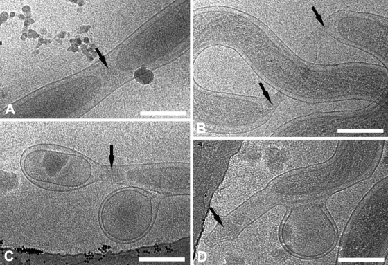
FIG. 7.
Events during cell division and the generation of cell envelope-derived blebs may help to explain the pleomorphism of T. pallidum ends. (A and B) Cryo-electron micrographs of early (A) and late (B) stages of division show nascent cone formation (black arrows). (C and D) Treponemes are shedding cell envelope-derived blebs which retain both outer and cytoplasmic membranes. Splotches are frost contamination. Bars, 200 nm.

