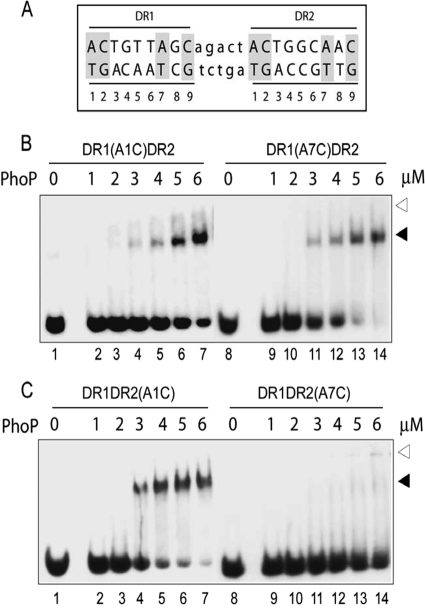
FIG. 2.
Roles of conserved adenines of DR1 and DR2 repeat motifs in PhoP-DNA interactions. (A) Sequence of the 23-bp core binding region comprising two direct repeat motifs (indicated in uppercase letters) with a 5-bp intervening spacer region (indicated in lowercase letters). The nucleotides in both repeat motifs are numbered from 5′ to 3′ (at the bottom), and the nucleotides conserved in all three direct repeat motifs are indicated by gray shadings. (B and C) EMSA of ∼20 nM oligonucleotide-based DNA probes with the indicated concentrations of PhoP using duplexes carrying substitutions at the A1 (lanes 1 to 7) and A7 (lanes 8 to 14) of the DR1 (B) or DR2 motifs (C). Direct-repeat specific substitutions are shown above each panel. The reaction condition is as described in Materials and Methods. Protein-DNA complexes were analyzed as described for Fig. 1A. The gels are representative of three independent experiments.

