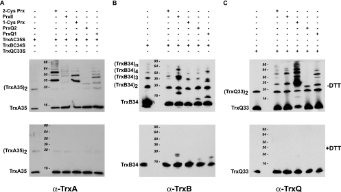
FIG. 2.
Analysis of the thioredoxin-peroxiredoxin interaction. Twenty (A) or 11.25 (B and C) μg of each peroxiredoxin was incubated with 8 μg TrxA(C35S) (A), 4 μg TrxB(C34S) (B), or 4 μg TrxQ(C33S) (C). Thereafter, proteins were resolved on 12% SDS-PAGE gels under reducing (lower panels) or nonreducing (upper panels) conditions. Thioredoxins were detected by Western blotting using specific antibodies at the following dilutions: anti-TrxA (α-TrxA), 1:1,000; α-TrxB, 1:10,000; α-TrxQ, 1:5,000.

