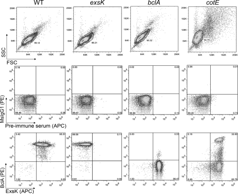
FIG. 3.
Two-color flow cytometric analysis of B. anthracis (Sterne) spores stained with anti-ExsK and anti-BclA antibodies. Wild-type (WT), exsK, bclA, and cotE spores were stained with anti-ExsK serum (allophycocyanin [APC]) and anti-BclA (phycoerythrin [PE]) antibodies. Top, FSC versus SSC dot plots. Middle, isotype control (mouse IgG1 [MsIgG1] and preimmune serum) dot plots. Bottom, anti-ExsK and anti-BclA antibody dot plots. Gates in each FSC versus SSC plot indicate the population used for isotype and antibody stain analyses.

